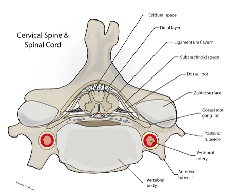
Subarachnoid Space Spine
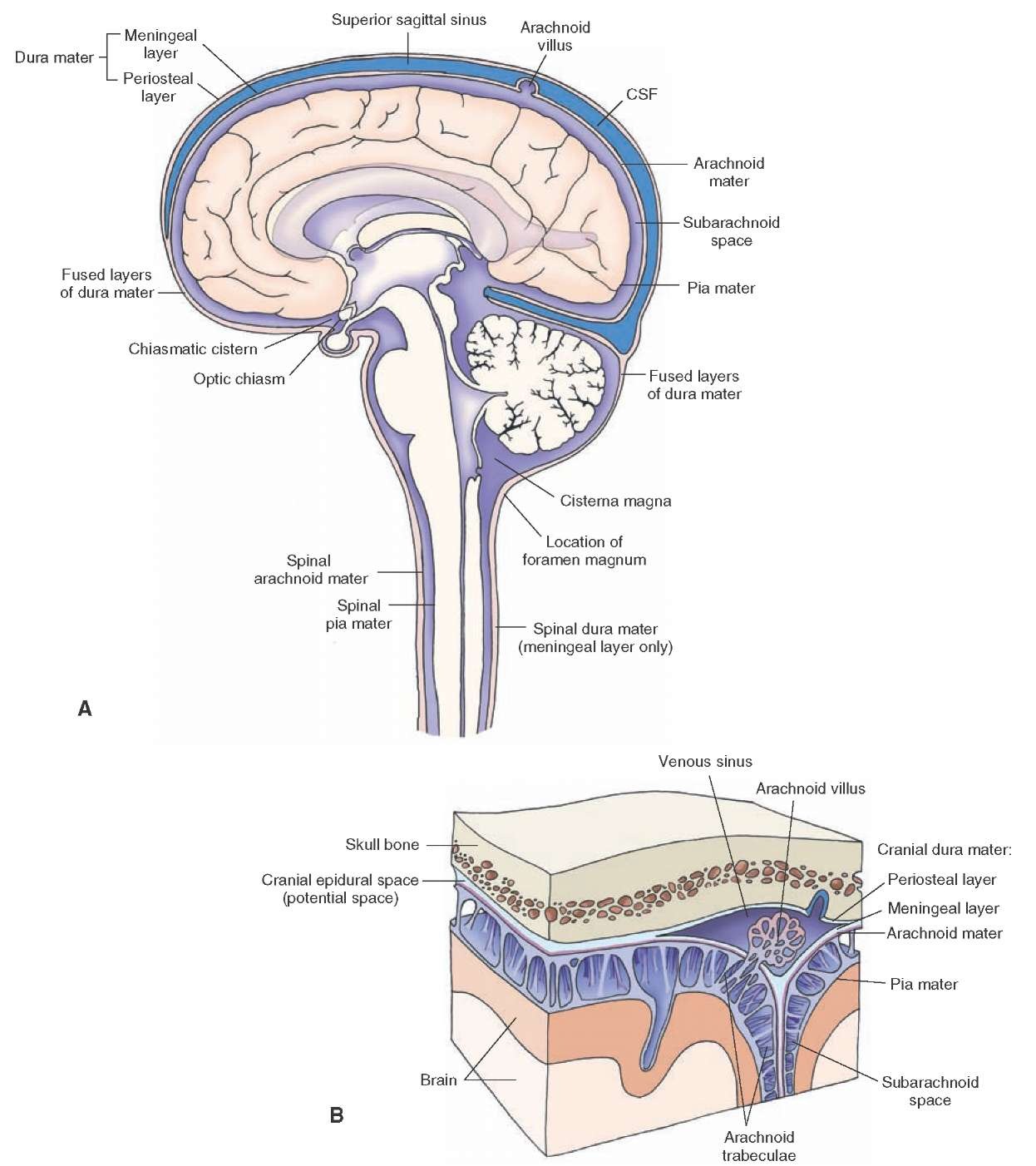
Meninges And Cerebrospinal Fluid Gross Anatomy Of The Brain Part 1
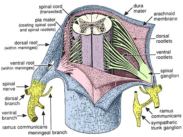
Lab 2 Spinal Meninges

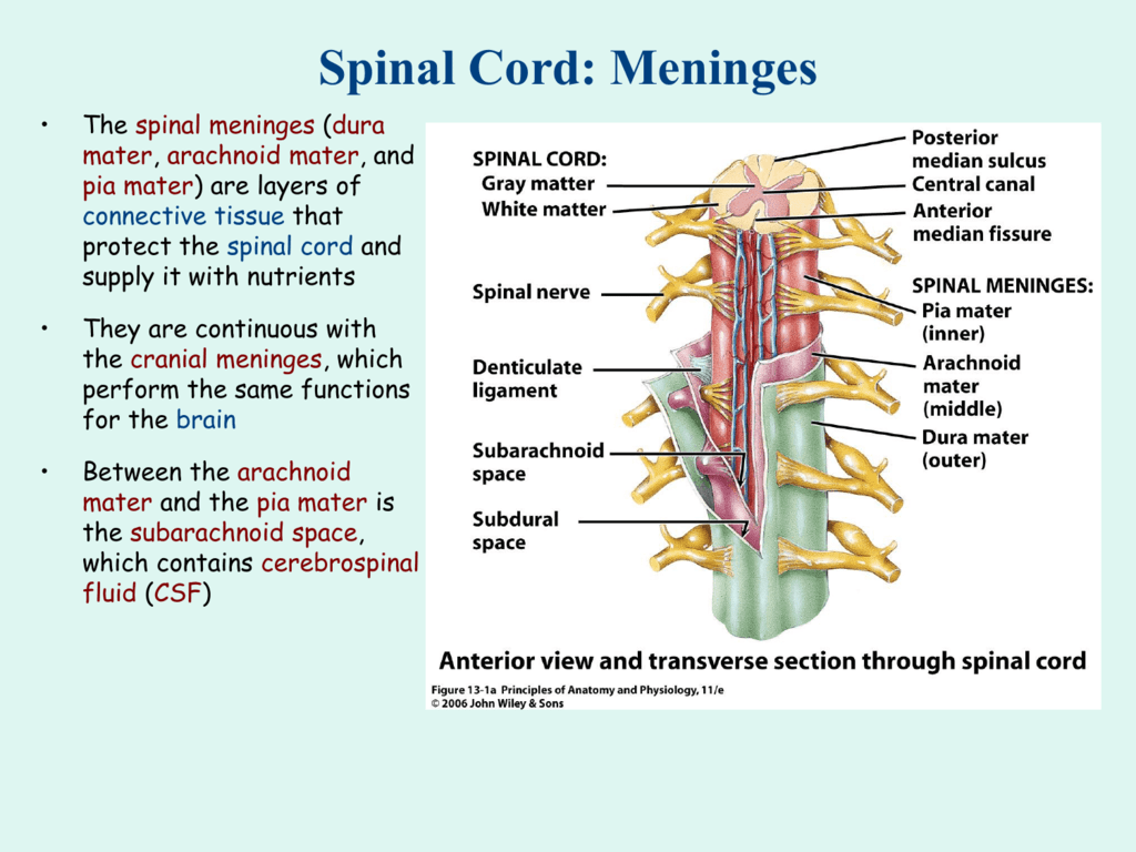
Spinal Cord Anatomy 1

Spinal Subarachnoid Space Google Search Spinal Cord Medical

Neuraxial Anatomy Nysora
Spinal Meninges

Extraventricular Origin Of The Cerebrospinal Fluid Formation Rate
Epi on upon dura mater also known as epidural cavity extradural space or peridural space.
Subarachnoid space spine. Spaces are formed from openings at different points along the subarachnoid space. The anatomy term epidural space has its origin in the ancient greek language. The subarachnoid space is the interval between the arachnoid membrane and the pia mater. 1120 15th street augusta ga 30912 5563 spine at west wheeler 706 869 1515.
Spinal anaesthesia or spinal anesthesia also called spinal block subarachnoid block intradural block and intrathecal block is a form of neuraxial regional anaesthesia involving the injection of a local anaesthetic or opioid into the subarachnoid space generally through a fine needle usually 9 cm 35 in longit is a safe and effective form of anesthesia performed by nurse anesthetists. It is occupied by delicate connective tissue trabeculae and intercommunicating channels containing cerebrospinal fluid csf. The discs in the spine that separate and cushion vertebrae may dry out and herniateas a result the space between the vertebrae shrinks and the discs lose their ability to act as shock absorbers. The dura mater is attached to the skull whereas in the.
The cavity is small in the normal brain. Spine glossary subarachnoid space. In humans the epidural space contains lymphatics spinal nerve roots loose connective tissue adipose tissue small arteries dural venous sinuses and a network of internal vertebral venous plexuses. In the central nervous system the subarachnoid space also called the subarachnoid cavity is the area in the brain between the arachnoid membrane which is the middle of three membranes covering the surface of the brain and the pia mater which is the deepest protective membrane covering the brain.
Over the gyri the arachnoid membrane and pia mater are in close contact. With this procedure the csf can be analyzed and measured a spinal anaethetic given or contrast be injected for myelography. Cervical stenosis occurs when the spinal canal narrows and compresses the spinal cord and is most frequently caused by aging. As the spinal cord ends at the level of l2 the subarachnoid space distal to this forms the lumbar cistern and is an ideal space to access the csf via a lumbar puncture 2.
The subarachnoid space is a substantial space between the arachnoid and the pia mater it contains the cerebrospinal fluid. The spinal cord 2009 related terms.
