Pleural Space Diagram
Lung Anatomy Saint Luke S Health System
Parietal Pleura

Pleural And Mediastinal Lung And Airway Disorders Merck

Pathology Glossary Pleural Effusions Draw It To Know It
Clinical Aspects Of Pleural Fluid Ph
Condition Specific Radiology Pleural Effusion Stepwards
Pleura Space Anatomy Charalampidis Journal Of Thoracic Disease
1 under normal conditions the pleural space contains 01 to 02 mlkg of fluid with a protein concentration of less than 15 gdl that flows down gravity dependent gradients.

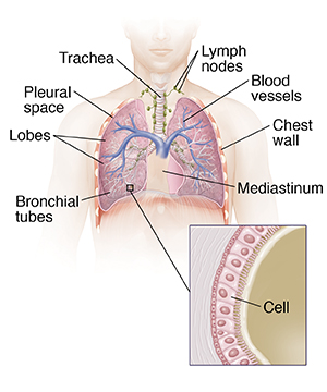
Pleural space diagram. The pleura is a serous membrane which folds back onto itself to form a two layered membrane structure. Pleura is formed by an inner visceral pleura and an outer parietal layer. When the lung collapses however or when air or liquid collects between the two membranes the pleural cavity or sac becomes. The outer pleura is attached to the chest wall.
Each pleural cavity is defined by a space surrounding each lung and is lined by a pleural membrane. Excluded from global pleural pressures by the anatomic location of the tube normal pleural healing or pathologic changes eg blood clots. These layers can be illustrated in a transverse. The thin space is known as the pleural cavity and contains a small amount of pleural fluid few milliliters in a normal human.
The pleural cavity is the potential space between the two pleurae visceral parietal of the lungs. The parietal pleura folds back on itself at the root of the lung to become the visceral pleura. In health the two pleurae are in contact. Schematic diagram of the pleural membranes cross section through thoracic cavity at the level of the heart each lung is placed within a separate layer of membrane thus there are two pleural sacs.
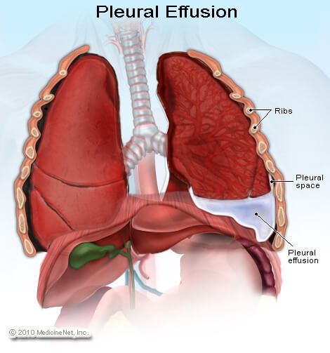
Expiratory air leak if the pleural tube is adequately draining the pleural space and there is no air accumulation the system will rapidlyreachequilibriumwiththeapplicationofxedsuc tion eg 20 cm h. The normal pleural space is approximately 18 mm wide at its least dependent point and widens to about 20 mm in the dependent regions. A pleural effusion is excess fluid that accumulates in the pleural cavity the fluid filled space that surrounds the lungsthis excess fluid can impair breathing by limiting the expansion of the lungs. Pleura plural pleurae or pleuras membrane lining the thoracic cavity parietal pleura and covering the lungs visceral pleura.
Various kinds of pleural effusion depending on the nature of the fluid and what caused its entry into the. The space between the two sacs is known as the mediastinum and is almost in the midline of the thorax. The pleural membrane is made up of two layers. The pleural cavity also known as the pleural space is the thin fluid filled space between the two pulmonary pleurae known as visceral and parietal of each lunga pleura is a serous membrane which folds back onto itself to form a two layered membranous pleural sac.
The outer pleura parietal pleura is attached to the chest wall but is separated from it by the endothoracic fascia. 23 the pleural membranes and the space they define play an integral function in.
