Intercostal Space Diagram

Intercostal Space Diagram Diagram Quizlet

12 Lead Ecg Placement Guide With Illustrations

2 Embryology Anatomy And Physiology Of The Lung Thoracic Key

Lung Surface Anatomy And Chest Tubes Vs Needle Decompression
Muscles Of Respiration Ppt Download

Chapman Reflex Points Flashcards Memorang
Arrow Showing Location For Needle Thoracocentesis In The 5th
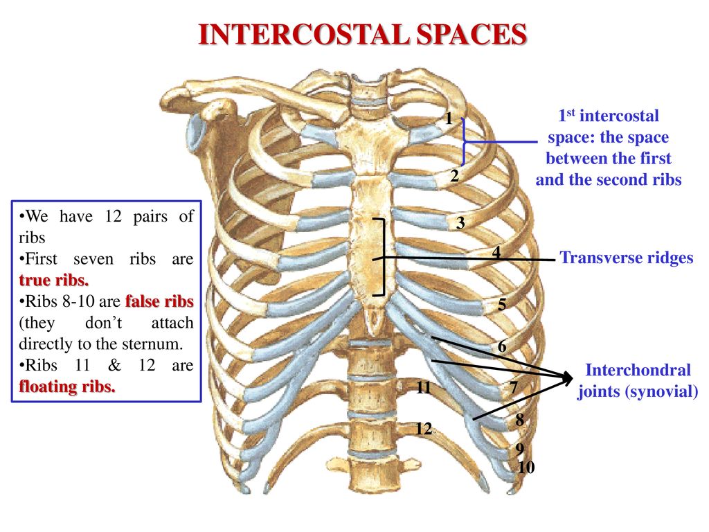
Start studying intercostal space.

Intercostal space diagram. Several kinds of intercostal muscle. Typical intercostal space lutfi abdul muien. Since there are 12 ribs on each side there are 11 intercostal spaces each numbered for the rib superior to it. 13 enumerate the factors responsible for the increase in various diameters of the thoracic cavity during inspiration.
Matthew grouthamel md reviewer. Unsubscribe from lutfi abdul muien. Each intercostal nerve enters the corresponding intercostal space between the posterior intercostal membrane and the parietal pleura. The nerve then travels forward with the intercostal vessels in the costal groove of the corresponding rib between the internal and innermost intercostal muscles.
Bordered by the rib above and below the deep fascia of the thorax superficially and the endothoracic fascia and pleura internally the intercostal. Intercostal muscles duration. Intercostal arteries and intercostal veins. Learn vocabulary terms and more with flashcards games and other study tools.
Chest ecg electrodes and placement. Spaces between the ribs are known as intercostal spaces. V1 fourth intercostal space on the right sternum v2 fourth intercostal space at the left sternum v3 midway between placement of v2 and v4 v4 fifth. Each intercostal space is lined on the inside by endothoracic fascia which is in turn lined with the parietal.
Carpal tunnel duration. The first thoracic nerve divides into a superior part which joins the brachial plexus and an. Structures in intercostal space. Diagram for intercostal space article updating please wait.
Each space contains three muscles of respiration namely the external intercostal muscle the internal intercostal muscle and the innermost intercostal muscle. 12 draw labelled diagram showing tributaries of azygous vein. May 15 2020 the eleven paired intercostal spaces contain the intercostal muscles nerves arteries veins and investing fascia. Dimitrios mytilinaios md phd last reviewed.
The intercostal space ics is the anatomic space between two ribs lat. 11 draw transverse section ts of intercostal space showing intercostal muscles and course branches of intercostal nerve. Every space consists of intercostal muscles and neurovascular bundle of intercostal plane. Easy anatomy 22748 views.
